TechBullion
3d
325

Image Credit: TechBullion
How Can Specular Microscopy Improve Eye Exams?
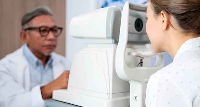
- The Specular Microscope is a transformative technology that enables ophthalmologists to examine the cornea of the eye at a cellular level.
- Specular microscope helps to examine the inner surface of the cornea and images it in great detail, helping doctors detect the early signs of a problem before other factors.
- Specular microscopy is a quick, painless, and non-invasive procedure that captures detailed images of the inside of the cornea, evaluating ear health by analyzing the density, size and shape of the cells.
- The biggest advantage of the specular microscope is that corneal diseases, such as Fuchs’ dystrophy, corneal edema, and keratoconus, can be detected early, normally before they begin to affect the vision.
- Using specular microscopes, doctors can diagnose and monitor several eye conditions that include Fuchs’ dystrophy, corneal edema, keratoconus, and corneal transplant rejection.
- This procedure helps post eye surgery, such as cataract removal or corneal transplant in keeping an eye on the cornea’s healing, ensuring that endothelial cells are not stressed.
- Using specular microscopes, the doctor can tailor a treatment plan as per the patient’s needs, whether managing corneal disease or tracking progress after surgery, offering personalized care with the best outcomes possible.
- Since specular microscopes give detailed images, doctors can use them to assess the health of the cornea when recommending eye treatment procedures, determining whether someone is a suitable candidate for surgery.
- Specular microscopes are quiet heroes of the world of eye care, working hard to keep your vision safe, and doctors believe that they will soon become part of every eye exam.
- Patients should not be worried when undergoing an examination with a specular microscope. It is a simple, painless, and quick test-taking high-resolution images of the eyes, helping the doctor detect problems early and providing the best preventative eye care.
Read Full Article
19 Likes
For uninterrupted reading, download the app