Physicsworld
2M
394

Image Credit: Physicsworld
Imaging method could detect Parkinson’s disease up to 20 years before symptoms appear
- Researchers at Tel Aviv University in Israel have developed a method to detect early signs of Parkinson’s disease at the cellular level using skin biopsies.
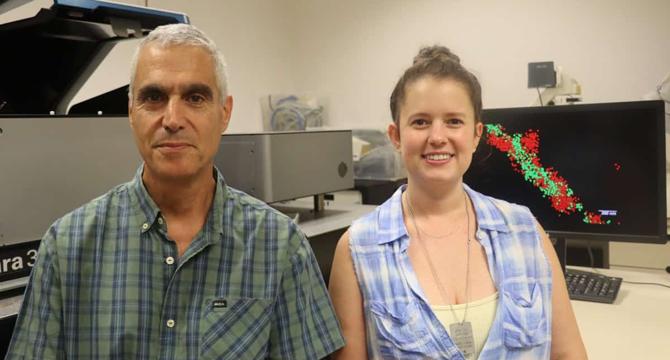
- The new method combines a super-resolution microscopy technique, known as direct stochastic optical reconstruction microscopy (dSTORM), with advanced computational analysis to identify and map the aggregation of alpha-synuclein (αSyn), a synaptic protein that regulates transmission in nerve terminals.
- The analysis detected a larger number of clusters, clusters with larger radii, and sparser clusters containing a smaller number of localizations in Parkinson’s disease patients relative to what was seen with healthy control subjects.
- Parkinson’s disease diagnosis based on quantitative parameters represents an unmet need that offers a route to revolutionize the way Parkinson’s disease and potentially other neurodegenerative diseases are diagnosed and treated.
Read Full Article
23 Likes
For uninterrupted reading, download the app